Lateral Scapula Radiography wikiRadiography
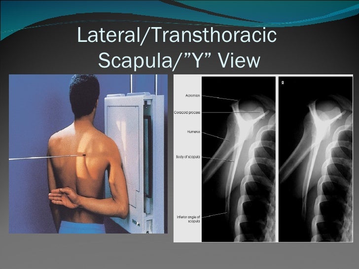
centering point. the level of the glenohumeral joint on the posterior aspect of the patient (5 cm below the top of the shoulder) central to the medial scapula border. collimation. laterally to include the skin margin. medially to cover the entirety of the medial scapula. superior to the skin margin. inferior to the inferior angle of the scapula.
Scapular Y View ALiEM
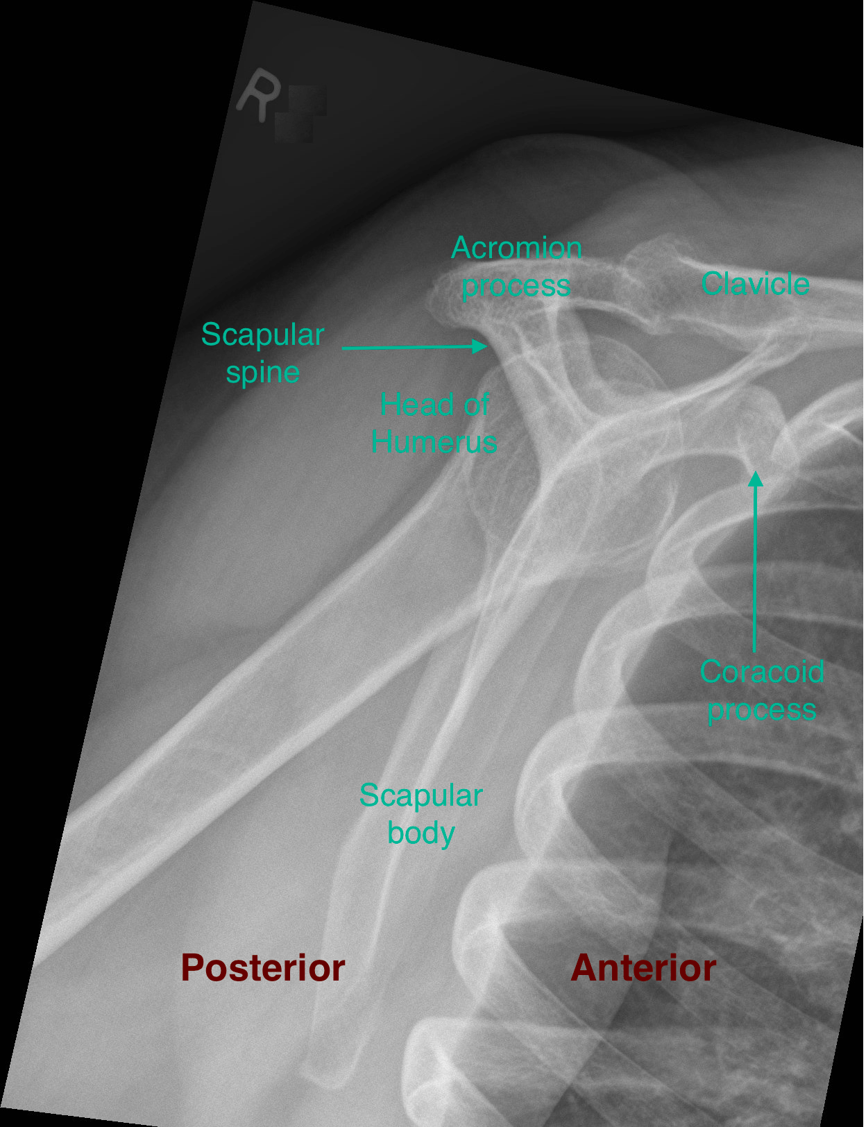
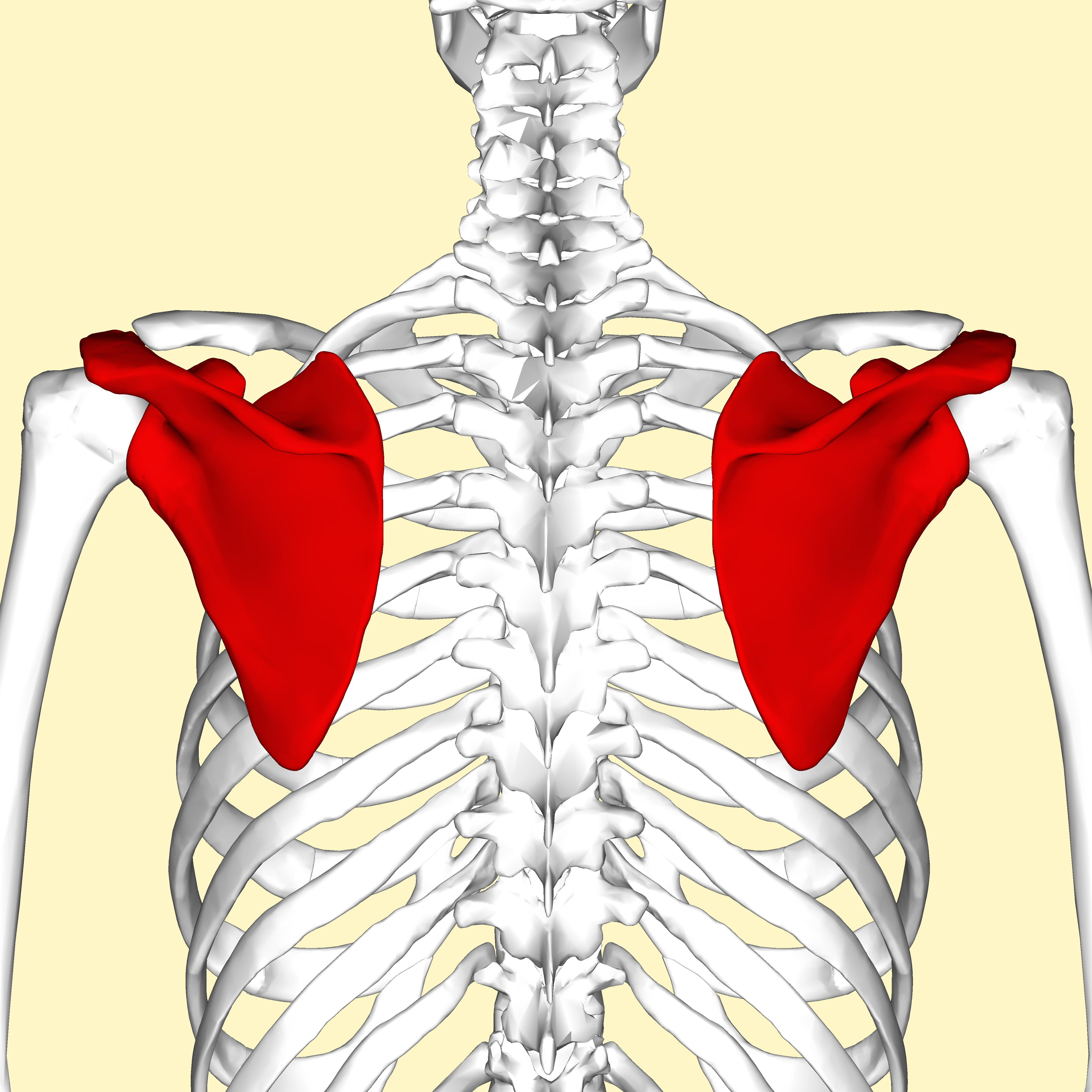
The scapula (or shoulder blade) is one of the two bones that form the pectoral girdle, the other being the clavicle. It's a large, triangular bone that is found along the posterior aspect of the upper thoracic cage. It's classified as a flat bone and includes the following bony features: - surfaces: costal and dorsal surfaces, and medial.
Proyeksi pemeriksaan Scapula Aditya Radiografer
Like any triangle, the scapula consists of three borders: superior, lateral and medial. The superior border is the shortest and thinnest border of the three. The medial border is a thin border and runs parallel to the vertebral column and is therefore often called the vertebral border. The lateral border is often called the axillary border as it runs superolaterally towards the apex of the axilla.
Scapula Stability My Family Physio
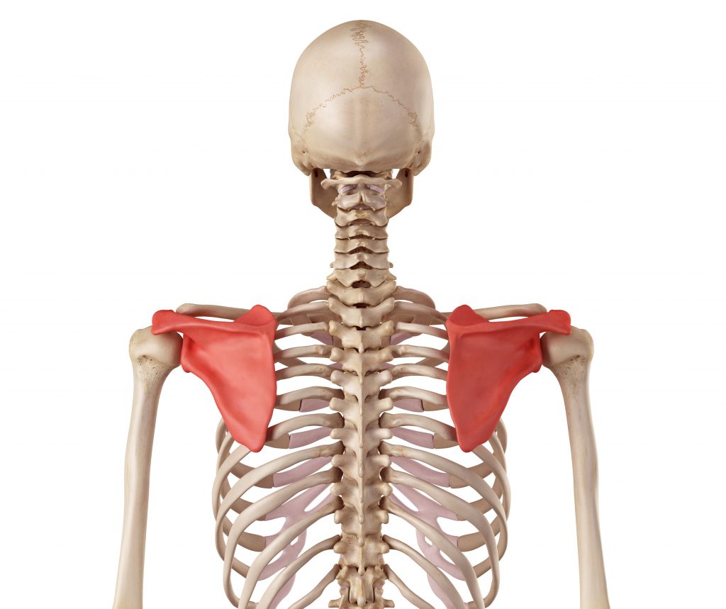
What is Scapula. The scapula, alternatively known as the shoulder blade, is a thin, flat, roughly triangular-shaped bone placed on either side of the upper back. This bone, along with the clavicle and the manubrium of the sternum, composes the pectoral (shoulder) girdle, connecting the upper limb of the appendicular skeleton to the axial skeleton.

Pin on Radiology
Pemeriksaan LGS skapula secara aktif dilakukan oleh pasien. Gerakan pada video ini meliputi:1. Elevasasi skapula (scapular elevation)2. Depresi skapula (scap.

Proyeksi pemeriksaan Scapula Aditya Radiografer
LAO (body of scapula) - 45°. LAO (Acromion) - 60°. Proyeksi : Lateral (Recumbent) - LPO/RPO. Kaset : ukuran 24 x 30 cm. kV : 75 ± 5 mAs :13. FFD : 100 cm. Posisi Pasien : Pasien supine di atas meja pemeriksaan dan posisikan lengan menyilang di depan dada. Kemudian rotasikan tubuh 30° atau sesuai kebutuhan agar scapula berada dalam.
Ppshoulder
A video tutorial that covers the structure and actions of the scapulothoracic joint.Access my FREE Online Membership today → https://www.thenotedanatomist.co.

Simulasi Pemeriksaan Radiologi (Thorax PA) YouTube
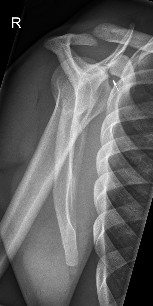
Citation, DOI, disclosures and article data. The scapula series is the plain radiographic assessment of the scapular bone of the shoulder girdle, seldom used in departments with 24 hour computed tomography departments. Many radiographic departments, do not have a stand alone scapula series, rather include the assessment of the scapula in the.

TEKNIK PEMERIKSAAN RADIOGRAFI OS SCAPULA Y VIEW LK PKL 1 YouTube
Scapular dyskinesis is the term used for abnormal function or mobility of your scapula. This can occur due to many things, including: Imbalance, tightness, weakness, and occasionally detachment of.
Proyeksi pemeriksaan Scapula Aditya Radiografer
The term 'winged scapula' (also scapula alata) is used when the muscles of the scapula are too weak or paralyzed, resulting in a limited ability to stabilize the scapula. As a result, the medial or lateral borders of the scapula protrudes from back, like wings. The main reasons for this condition are musculoskeletal- and neurological-related.
Scapula Ten Gates to Heaven
The posterior surface of the scapula (or shoulder blade) has a prominent ridge of bone know as the spine of scapula. It is a shelf-like projection that separates the posterior surface of the scapula into two parts: the superior supraspinous fossa and the inferior infraspinous fossa. The spine of scapula can be readily palpated and serves as an.

Scapula Fracture ALiEM
Proyeksi Pemeriksaan Lateral. Kriteria gambaran : Scapula, Coracoid Process, Acromion, Inferior angle. PP (Posisi pasien) = Pasien berdiri (Erect) membelakangi arah sinar PO (Posisi Objek) = Siku pada sisi yang diperiksa dalam keadaan fleksi, lengan sedikit abduksi dan diletakkan dibelakang tubuh dan tubuh dirotasikan 60-70 derajat sehingga.

An AP radiograph of the left scapula shows a lucent, expansile and... Download Scientific Diagram
A commonly observed abnormal pattern of scapular motion (scapular dyskinesis) is the premature or excessive scapular elevation that appears as shrugging ( Fig. 93-3 ). This pattern has been associated with rotator cuff pain, weakness, and fatigue. It has also been observed with loss of glenohumeral motion.
Proyeksi pemeriksaan Scapula Aditya Radiografer
The scapula or shoulder blade is the bone that connects the clavicle to the humerus. The scapula forms the posterior of the shoulder girdle. It is a sturdy, flat, triangular bone. The scapula provides attachment to several groups of muscles. The intrinsic muscles of the scapula include the rotator cuff muscles, teres major, subscapularis, teres minor, and infraspinatus. These muscles attach.

winging scapula test YouTube
The scapula is described as having superior, medial, and lateral borders. Posteriorly, the scapula is divided into a supraspinous fossa and infraspinous fossa by the scapular spine. Anteriorly, on the costal surface, is the shallow subscapular fossa. Laterally is the glenoid fossa, anteriorly is the coracoid process and superiorly is the.
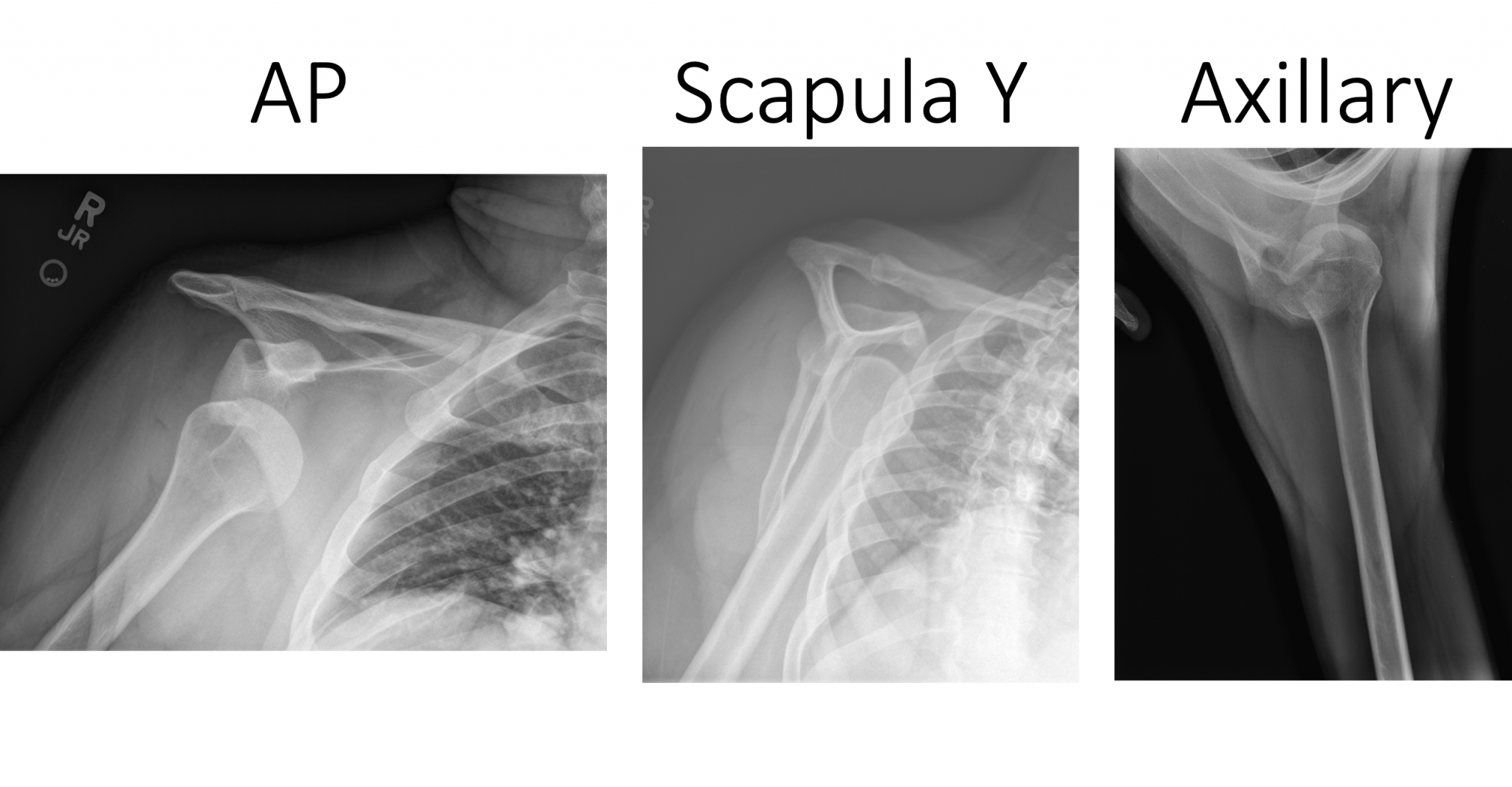
Emergency Medicine EducationEM3AM Anterior Shoulder Dislocation
Teknik Pemeriksaan. Proyeksi AP Posisi Pasien : Pasien diposisikan erect atau supine. Posisi Objek : - Scapula menempel kaset - Lengan diangkat ke atas kepala - atur scapula pada pada pertengahan pasien. Kriteria Radiograf : - lateral dan medial scapula saling superposisi